Wednesday, November 10, 2010

Lecture - Molecular techniques (nucleic acids)

In todays lecture we focused on techniques that are used for analyzing nucleic acids. I decided to focus mainly on DNA sequencing, given how important that such technique has become in the last decade. Topics that were discussed:
  • PCR
  • PCR in disease diagnosis
  • DNA sequencing
    • Chain termination method (Sanger method - manual and automated)
    • Shotgun sequencing
    • Pyrosequencing
    • Next-generation sequencing
  • DNA typing
----------------------

Tuesday, November 9, 2010

Lecture - Molecular techniques (proteins)

In this lecture we had a snapshot of what could easily be a whole course just on molecular techniques. We focused on protein analysis techniques:
  • Purification (column chromatography)
  • Separation (SDS PAGE and two-dimensional electrophoresis)
  • Detection (western blotting)
  • Predicting function (using bioinformatic tools)
We also introduced the basics of techniques focused on nucleic acids, including a discussion of the constantly decreasing cost of sequencing complete genomes.

----------------------

Thursday, November 4, 2010

Module 4, Lab 13 - Links for Bioinformatics lab 1

----------------------

Module 3, Lab 12 - RED of pGLO plasmid

Today we did a restriction enzyme digestion (RED) of the plasmid DNA purifications (minipreps) we did last week. The goal was to isolate the GFP gene from the pGLO plasmid. After the cloning process that would have been a step before purifying the GFP gene sample for further study.

We used the restriction enzymes EcoRI and HindIII (individually and in combination) to reach our goal. We confirmed the results with an agarose gel electrophoresis (students in section 2 even did a "retrophoresis"... Hopefully it was a very valuable lesson)

----------------------

Tuesday, November 2, 2010

Lecture, chapter 11 - Gene regulation at the mRNA level

Today we covered chapter 11, on gene regulation at the mRNA level. Regulation mechanisms in the middle ground between transcriptional regulation and translational regulation.

We discussed the control of rate of degradation of mRNA, the effect of translational regulatory proteins (activators and/or repressors), regulation by anti-sense RNA, and regulation by alterations to the ribosome.

Tomorrow we will discuss RNA interference (RNAi)

----------------------

Saturday, October 30, 2010

Module 3, Lab 11 - PAGEs of GFP

Image from Bio Rad

As a follow up of Thursday's lab, we stained the gels with Coomasie G-250 stain and then air dried them for analysis.


(click on pic for a full size image)

-----------------------

Module 3, Lab 12 - Small Scale Plasmid DNA Purification of pGLO

In this lab students purified the pGLO plasmid following Promega's Wizard Plus SV Minipreps DNA Purification System®.

Next week we will do a restriction enzyme digestion of the plasmid DNA in order to isolate the GFP gene from the plasmid.

----------------------

Thursday, October 28, 2010

Module 3, Lab 11 - Native and denaturing polyacrylamide gel electrophoreses (PAGEs) of GFP

Today we ran a new kind of electrophoresis: Polyacrylamide Gel Electrophoresis (PAGE) is a process that uses the same principle of agarose gel electrophoresis, but it uses a polyacrylamide gel, a thinner, more expensive kind of gel that provides a higher resolution than its agarose counterpart.

We specifically ran protein samples in two ways.
  • A native gel, in which the proteins, in their native state, migrate at different rates depending on their size (molecular weight), 3D structure, and charge.
  • A denaturing gel, in which the proteins are denatured (linearized) in the presence of a detergent such as Sodium Dodecyl Sulfate (SDS) that coats the proteins with a negative charge. The resulting denatured proteins have an overall negative charge and a similar charge to mass ratio. Since denatured proteins act like long rods instead of having a complex tertiary shape, the rate at which they migrate in the gel depends only to their size (molecular weight) and not its charge or shape.
The goal was to estimate the size of the green fluorescent protein (GFP) by comparing its migration through each gel with the migration of a molecular weight ruler (a "protein ladder") loaded onto the same gel.

PAGE is used for separating proteins ranging in size from 5 to 2,000 kDa due to the uniform pore size provided by the polyacrylamide gel. Agarose gels can also be used to separate proteins, but they do not have a uniform pore size, so they are optimal only for electrophoresis of proteins that are larger than 200 kDa.

We will be able to compare teh results in both gels, and if GFP has any activity in either one of them (through pictures taken under UV light).
----------------------

Wednesday, October 27, 2010

Exam 2

Statistics for exam 2

Click on pic for full size image

-----------------------

Friday, October 22, 2010

Module 3, Lab 10 - Protein quantitation
Bradford dtermination of GFP


Today we did a protein quantitation using BioRad's Quick Start™ Bradford Protein Assay, a method in which a dye reagent is used (Bradford reagent, based on Brilliant Blue G-250) to bind to proteins (causing the dye reagent to change from a reddish-brownish color to blue) and measure its absorbance. The more concentrated the protein it binds, the darker the blue resultant color, and the greater the absorbance at 595 nm.

Two relative standard proteins are used, bovine serum albumin (BSA) and bovine gamma-globulin (BGG), to generate absorbance vs. protein concentration curves and then interpolate the absorbance of problem samples (mostly with GFP) to estimate their concentration. The problem samples were obtained from the Hydrophobic Interaction Chromatography (HIC).

This method is applied when researchers in proteomics discover a new protein and are trying to gather information about it. In our case, we "discovered" GFP, although we wouldn't have a name yet, had it been a truly newly discovered protein.
-----------------------

Module 3, Lab 09 - Hydrophobic Interaction Chromatography (HIC) of GFP

Friday, October 21, 2010

Today we performed a Hydrophobic Interaction Chromatography (HIC) to separate the green fluorescent protein (GFP), produced in our bacterial cultures, from other proteins commonly found in bacteria.

A sample of bacteria was concentrated and then resuspended in a solution in which they were lysed. The high salt solution, containing all the proteins found in the bacteria, was then passed through a hydrophobic interaction column where molecules of GFP bound to the hydrophobic beads. The high salt solution increased the hydrophobicity of GFP by further exposing its hydrophobic amino acid residues.

A series of washes with buffers of decreasing salinity allows proteins with various levels of hydrophobicity to gradually unbind from the beads and be collected in a test tube. By switching collection tubes each time a buffer is added, different proteins can be collected. One of them was GFP and the tube in which it was collected should glow.

Diagram of Hydrophobic Interaction Chromatography (HIC)
GFP molecules are represented by black triangles
-----------------------

Module 2, Lab 07 - Sequencing reactions of GAPC gene

After the cloning process of the GAPC gene from Arabidopsis, and extracting the plasmid DNA (to isolate the pJet1.2 plasmid) we did a RED to confirm the success of the ligation.

Using the samples that had the GAPC gene insert we mixed purified plasmid DNA with forward and reverse sequencing primers (pJET SEQ F and pJET SEQ R), and put them in a 96-well plate. The plate will be shipped to the DOE Joint Genome Institute (JGI) to be sequenced as part of their Sequencing Training Program (STR). The results should be in in two weeks, ready to be used in the bioinformatics labs

We will discuss the DNA sequencing technique most commonly used: Dye-terminator sequencing, a modification of Sanger's chain termination sequencing protocol, which allowed the automation of the DNA sequencing process.
-----------------------

Wednesday, October 20, 2010

Guest lecture by Dr. Renee Reijo-Pera
Early human embryo development and associated gene expression

We had the privilege of having Dr. Renee Reijo-Pera, the director of the Human Embryonic Stem Cell Research Center at Stanford University, as a guest lecturer in our class. She shared with us her lab's findings in recent years on stem cell research and early human embryo development.

Some of the main topics in her lecture included...
  • Maternal vs. embryonic gene expression - Stages at which maternal mRNAs are active, and then degraded, and at which embryonic mRNAs are synthesized
  • Dynamics of cell division between fertilization and blastocyst stage
  • Prediction, at day 2 of development, of which embryos are viable (will successfully reach blastocyst stage) - Development of an algorithm to make an objective prediction
  • Things we do not know about human embryo development and how stem cell research can help
  • How embryo images were obtained and made into movies to allow analysis of developmental process
  • Analysis of gene expression - analysis of mRNA from 96 selected genes, extracted from a single cell
  • How the development process is correlated with patterns of gene expression
-----------------------

Tuesday, October 19, 2010

Lecture, chapter 11 - RNA processing
+ info about guest lecturer, Dr. Renee Reijo-Pera

Today we finished chapter 11 on RNA processing. We focused mainly on alternative splicing and how it produces different mRNA molecules by transcribing the same gene.


We also discussed processes like base modification, base substitution, RNA editing, and RNA degradation.


After finishing the chapter we discussed students' impressions on Dr. Reijo-Pera's Keiser lecture yesterday evening and expectations for her talk in our class tomorrow...!!! (expectations from the talk itself and about students' interaction with Dr. Reijo-Pera)


Tomorrow:


Dr. Renee Reijo-Pera, from Stanford University, will give a lecture on human preimplantation development and gene expression and pathways during the first few days of development. We will be joined by students in Dr. Aulthouse's Developmental Anatomy class, and potentially Dr. Walden's CLS program so the room will be packed. The talk will be as exciting as the Keiser lecture and having two-three classes in the audience will make the discussion more interesting and lively...!


Students should be ready to ask questions to, and engage in a discussion with, Dr. Reijo-Pera. Please check the following links:

It will be an exciting day. Take advantage of it!


-----------------------

Saturday, October 16, 2010

Upcoming lecturer: Dr. Renee Reijo-Pera


On Wednesday we will have the honor of having Dr. Renne Reijo-Pera as a guest lecturer in our class. Dr. Reijo Pera is visiting ONU as the Distinguished Keiser Lecturer of 2010.
Her lecture is titled Human Health, Development and Stem Cells, and it will be delivered on Monday, October 18, at 7:00 pm at the Freed Center

In our class Dr. Reijo-Pera will be talking about human preimplantation development and gene expression and pathways during the first few days of development. This topic is highly appropriate for our class and it will provide you with the opportunity to interact in an intimate setting with a world-class researcher. Take advantage of such opportunity!

Dr. Reijo-Pera is the Director of the Human Embryonic Stem Cell Research and Education Center at Stanford University. For more information see her lab web page, which includes information about her and her group, and her publications.

-----------------------

Module 2, Lab 06 - Cloning - RED of the plasmid DNApurifications

(entry in progress)
----------------------

Thursday, October 14, 2010

Module 2, Lab 06 - Cloning (GAPC gene from Arabidopsis and pJet1.2 plasmid)

Today we used the E. coli cultures you inoculated yesterday to do a small-scale plasmid DNA extraction ('miniprep') using Promega's Wizard® Plus SV Minipreps DNA Purification System.

Here's a recap of what we have done so far in the last couple of exercises:

Lab 5 - Nested PCR of the GAPC gene from Arabidopsis

Lab 6 - Cloning
  • Ligation of PCR amplified GAPC gene onto the pJet1.2 plasmid
  • Genetic transformation of E. coli with the pJet1.2 plasmid
  • Cloning of genetically transformed E. coli
  • Minipreps (purification of pJet1.2 plasmid)
Next, a restriction enzyme digestion to confirm successful plasmid DNA purification (lab 6) and setting sequencing reactions (lab 7)

-----------------------

Module 3, Lab 08 - Genetic transformation of E. coli with the pGLO plasmid

Jen was kind enough to prepare new LB agar plates and redo the transformation of E. coli with the pGLO plasmid. Then she plated the transformed bacteria on the plates she prepared, some of which had ampicillin, arabinose, or both.

[Fantastic] Results are shown in the pictures below:

(entry in progress)
----------------------

Wednesday, October 13, 2010

Lecture, chapter 11 - RNA processing


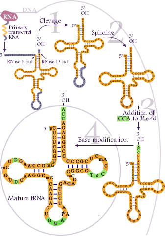
tRNA processing
------------------------------------------------------------------------------

Today we started chapter 11, on RNA processing, the fifth "stop" in our roadmap.

We mentioned the most basic processes through which RNA is modified (base modification, cleavage, and splicing) and described the more complex processes that are specific to eukaryotic RNA (5' capping and 3' polyadenylation).

We discussed in more detail the steps through which introns are spliced out the pre-mRNA molecule, and introduced the concept of alternative splicing.

-----------------------

Tuesday, October 12, 2010

Lecture, chapter 10 - Gene regulation in eukaryotes

Today we covered chapter 10, on gene regulation in eukaryotes.

We highlighted the differences in between the regulatory processes in prokaryotes and the more complex processes in eukaryotes, mainly the facts that in eukaryotes many transcription factors are needed to aid transcription and that DNA is many times condensed in heterochromatin, and therefore unavailable for RNA polymerase and other proteins.

Among some of the processes that affect gene expression we discussed histone acetylation, a mechanism through which heterochromatin is relaxed into euchromatin, making DNA available for DNA binding proteins, and methylation, a mechanism through which genes can be repressed or silenced, in some cases because it promotes de-acetylation of histones and DNA condensation into heterochromatin.
---------------------